Sense-by-growth (see 'Shaping with water') is one of the leading hypotheses for root perception of moisture. The theory posits that the uptake of water by growing root cells can serve as a readout for water availability in the environment. For example: cells in contact with a wet surface are able to grow quickly and 'easily' while cells in a soil air-gap may have more trouble sourcing the water they need to elongate. So far, the evidence for this gradient in 'ease of growth' across the root axis is computational. In this project, you'll see how I am trying to test the model with experiments.
Testing the computational sense-by-growth model requires a quantitative readout of cell state. In short, we want to build a reporter that can track the osmotic state of cells. For our use case, we want this reporter to be genetically-encoded, ratiometric, and responsive to hydration state without interfering with the delicate biology of the cell. The published Aquadust sensor was a major breakthrough in non-destructive measurements of water potential but only functions on surface tissue and falls short when it comes to internal cell layers.
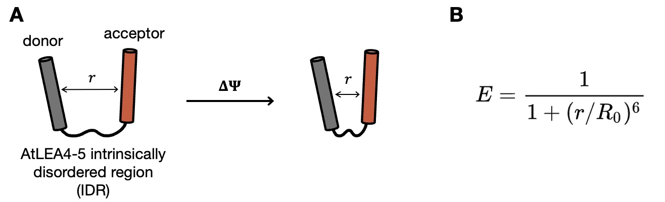
A) SED1 FRET sensor architecture as published in Cuevas-Velazquez, 2021. SED1 comprises two fluorophores: a donor, which is excited by incident light and can transfer this energy non-radiatively, and an acceptor, which emits light only when excited by the donor molecule. The IDR (intrinsically disordered) linker is the crux of the sensor. Upon some change in water potential ΔΨ, the linker condenses and brings the fluorophores closer together, which influences the aforementioned energy transfer. B) The amount of energy transferred (E) is highly sensitive to even minute changes in distance (r).'
Luck is on our side: we have a very nice starting point to design our sensor. The SED1 sensor, short for SENSOR EXPRESSING DISORDERED PROTEIN 1, was designed and tested in the Dinneny lab in 2021 as a reporter of molecular crowding, which is one of the physical consequences of a change in cell volume. SED1 combines the efficiency of Förster resonance energy transfer between two fluorophores with the sensitivity of intrinsically disordered regions (IDRs) to translate changes in cell state into visible optical signals that can be measured by microscopy. The SED1 sensor was shown to work beautifully in yeast cells and tobacco leaves but stubbornly refused to function in stable Arabidopsis transformants, which is arguably the most important place for it to work. This is where I come in.
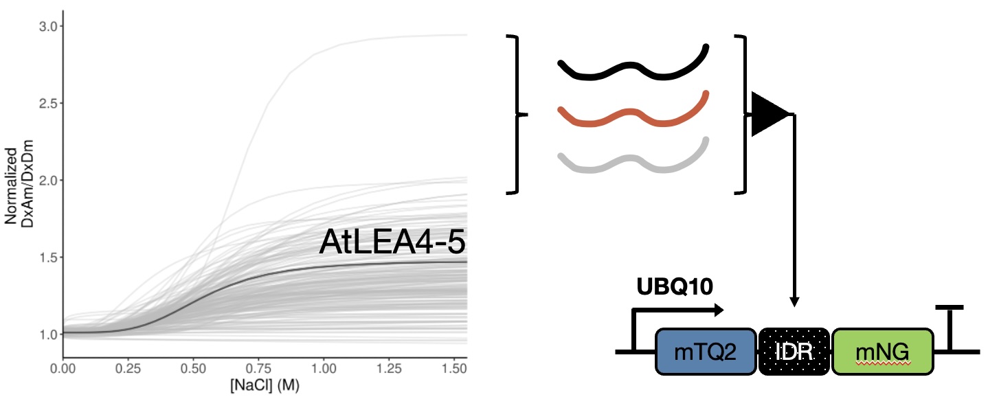
A panel of 200 IDRs sourced from different kingdoms of life was screened for sensitivity to NaCl. In total, 62 IDRs were measured as more sensitive than AtLEA4-5.
Cesar Cuevas-Velazquez, the original father of SED1, was kind enough to leave me with a list of IDRs he found were more sensitive than the original LEA4-5 linker in the SED1 sensor. I selected 10 of these candidates to swap into the SED1 architecture. Among them is a tardigrade protein known to vitrify under certain conditions; perhaps a mechanism for these amazing little creatures to survive prolonged states of extreme desiccation! To test the sensitivity of my 10 new SED variants, I transiently infiltrated them into tobacco leaves and measured fluorescence during salt or sorbitol treatment.
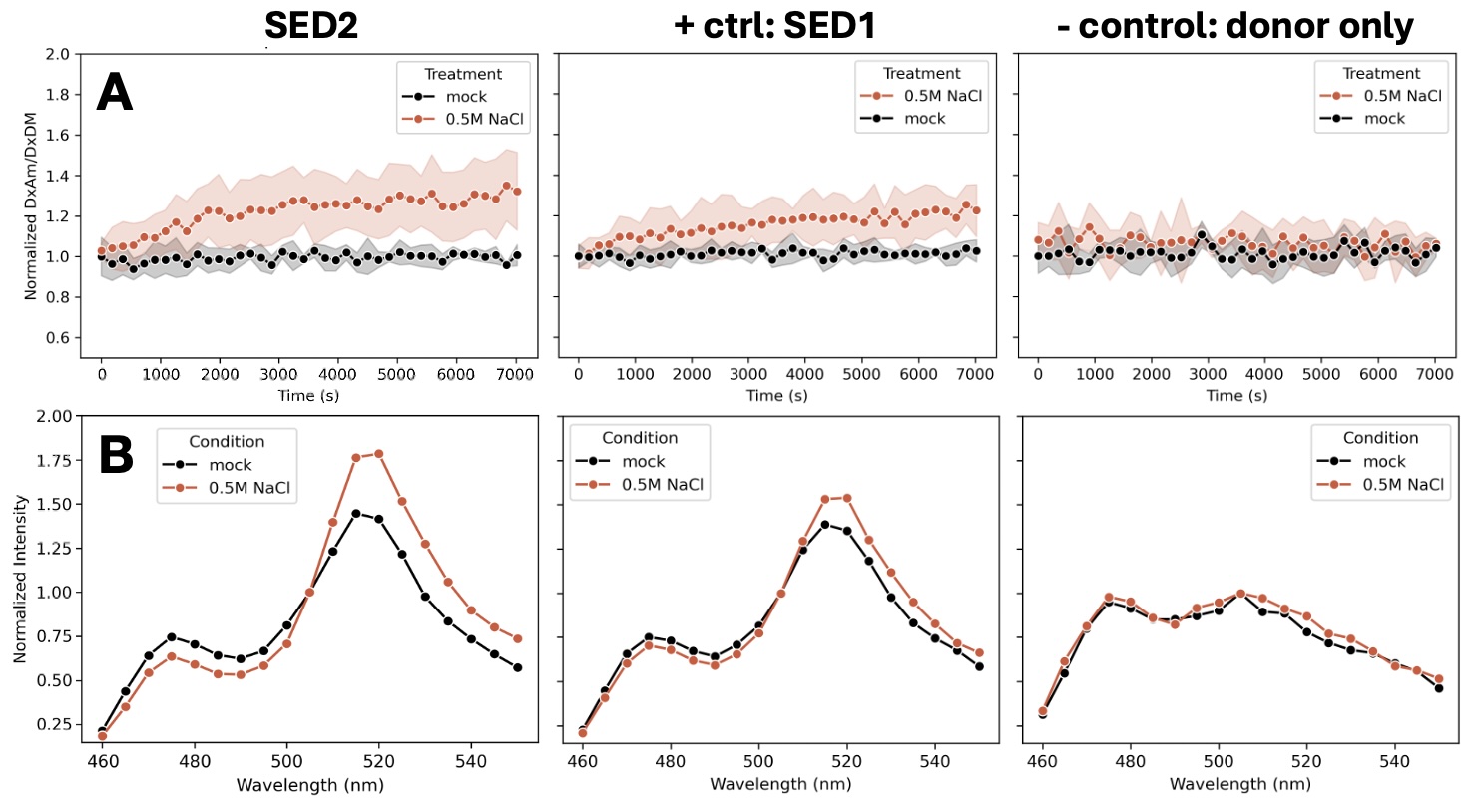
Sensor testing under transient expression in Nicotiana benthamiana (tobacco) leaves. A) Ratiometric FRET measurements (acceptor/donor fluorescence emission) under 434nm excitation; B) fluorescence emission spectra collected with 434nm excitation.
Several of the new SED variants tested displayed sensitivity on par with or greater than SED1 in tobacco leaf discs. Those variants were transformed into Arabidopsis thaliana and will be tested for sensitivity to salt- and osmoticum-treatment in the following months. Until then, suspense... A genetically encoded sensor of this sort would find many applications in the field of plant biology. Currently, the sensors are cytosol-localized, but future versions could target different organelles for specific applications.
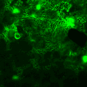
The localization patterns of leaf-infiltrated transgenes is often quite beautiful. In the above GIF, we see the typical puzzle-shaped cells of the leaf epidermis. Strong constitutive expression of SED2 drives localization in the cytosol, nuclei, and perhaps even cytoskeletal elements.
References
- Robbins NE II, Dinneny JR. (2018). Growth is required for perception of water availability to pattern root branches in plants. PNAS 115(4).
- Cuevas-Velazquez CL et al. (2021). Intrinsically disordered protein biosensor tracks the physical-chemical effects of osmotic stress on cells. Nat Commun 12.
- Jain P et al. (2021). A minimally disruptive method for measuring water potential in planta using hydrogel nanoreporters. PNAS 118(23):e2008276118.